Location, Dissection, and Analysis of the Murine Stellate Ganglion
Science never sleeps. Certainly not during the holidays. My wife has just published a new paper on the autonomic nervous system and its role with respect to cardiac electrophysiology.
→ Location, Dissection, and Analysis of the Murine Stellate Ganglion, Scherschel, Bräuninger et al., JoVE
The autonomic nervous system is a substantial driver of cardiac electrophysiology. Especially the role of its sympathetic branch is an ongoing matter of investigation in the pathophysiology of ventricular arrhythmias (VA). Neurons in the stellate ganglia (SG) – bilateral star-shaped structures of the sympathetic chain – are an important component of the sympathetic infrastructure.
The SG are a recognized target for treatment via cardiac sympathetic denervation in patients with therapy-refractory VA. While neuronal remodeling and glial activation in the SG have been described in patients with VA, the underlying cellular and molecular processes that potentially precede the onset of arrhythmia are only insufficiently understood and should be elucidated to improve autonomic modulation. Mouse models allow us to study sympathetic neuronal remodeling, but identification of the murine SG is challenging for the inexperienced investigator. Thus, in-depth cellular and molecular biological studies of the murine SG are lacking for many common cardiac diseases.
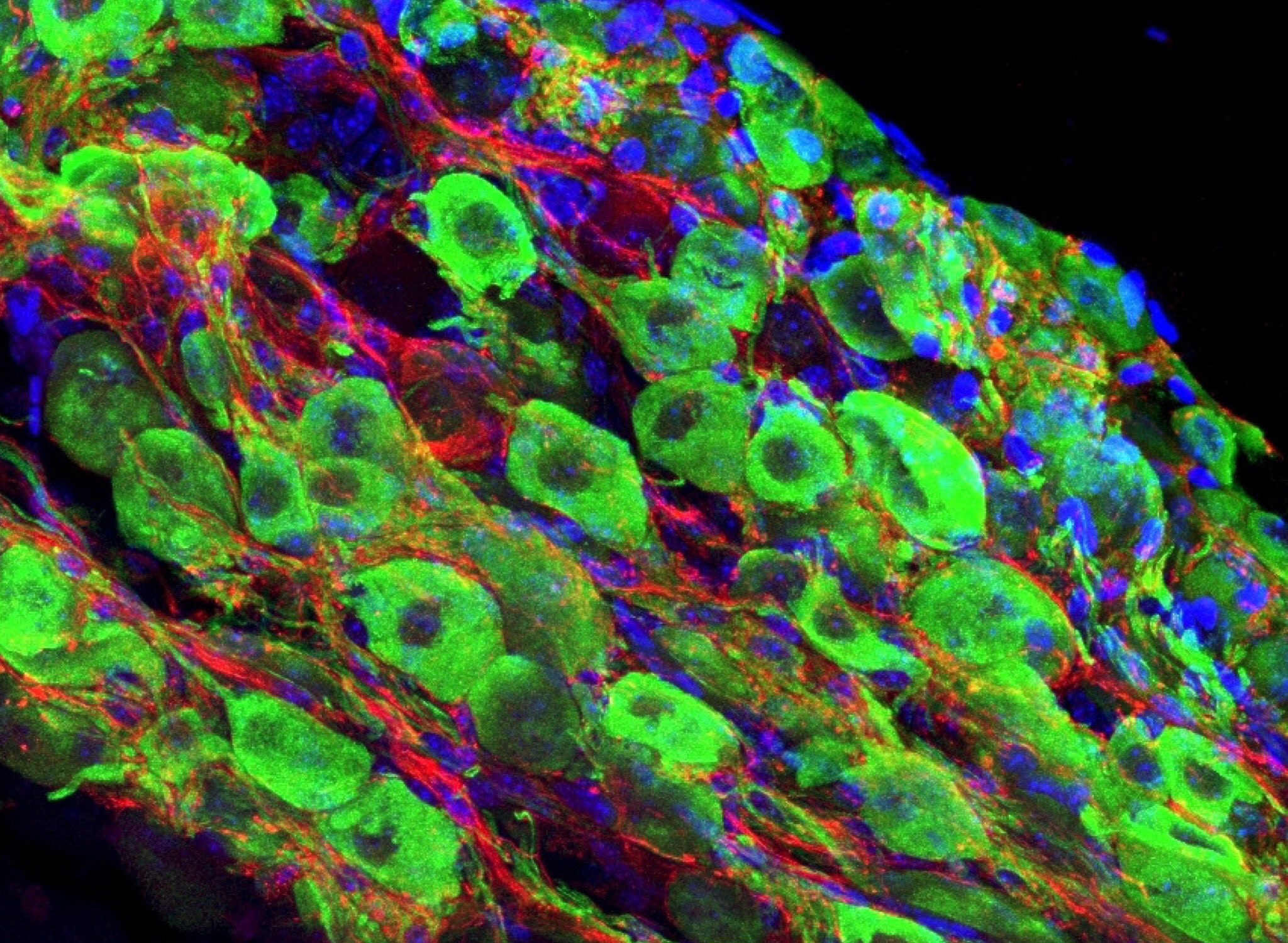
Here, we describe a basic repertoire for dissecting and studying the SG in adult mice for analyses at RNA level (RNA isolation for gene expression analyses, in situ hybridization), protein level (immunofluorescent whole mount staining), and cellular level (basic morphology, cell size measurement). We present potential solutions to overcome challenges in the preparation technique, and how to improve staining via quenching of autofluorescence. This allows for the visualization of neurons as well as glial cells via established markers in order to determine cell composition and remodeling processes. The methods presented here allow characterizing the SG to gain further information on autonomic dysfunction in mice prone to VA and can be complemented by additional techniques investigating neuronal and glial components of the autonomic nervous system in the heart.
Since JoVE is a video journal, there soon will be a video version of the paper. Which should have some nice visuals. Just look at this beautiful microscopy!